Gradual Worsening Upper and Lower Extremity Weakness
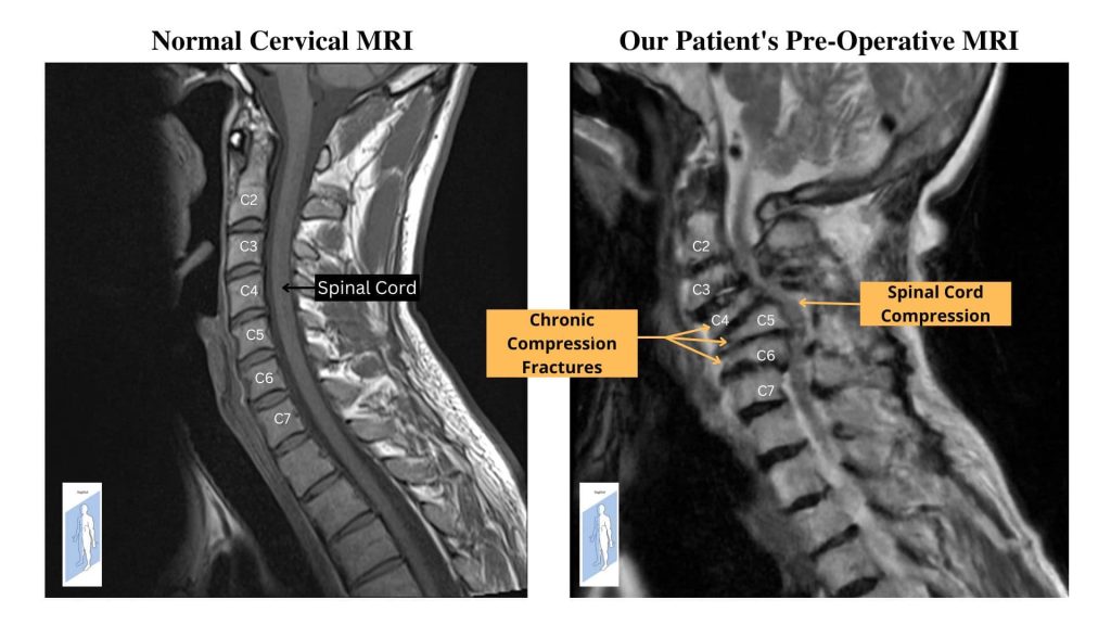
A 65-year-old female presented to the emergency department with complaints of gradual, worsening upper and lower extremity weakness. The patient’s weakness had progressed to the point where she was unable to complete daily activities or walk. MRI imaging revealed severe cervical disease including multiple, chronic compression fractures, severe spinal cord stenosis, and nerve compression (Picture #1.) Dr. Munshi was consulted and recommended to proceed with an anterior C4-6 corpectomy and posterior decompression and fusion of C2-7.
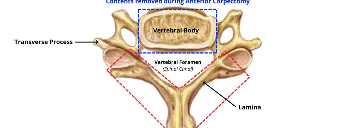
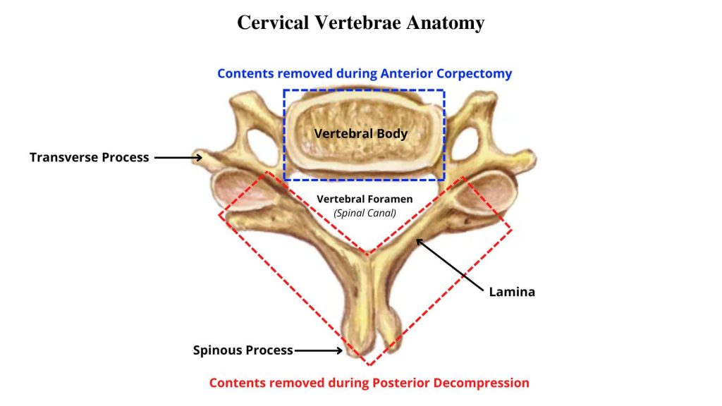
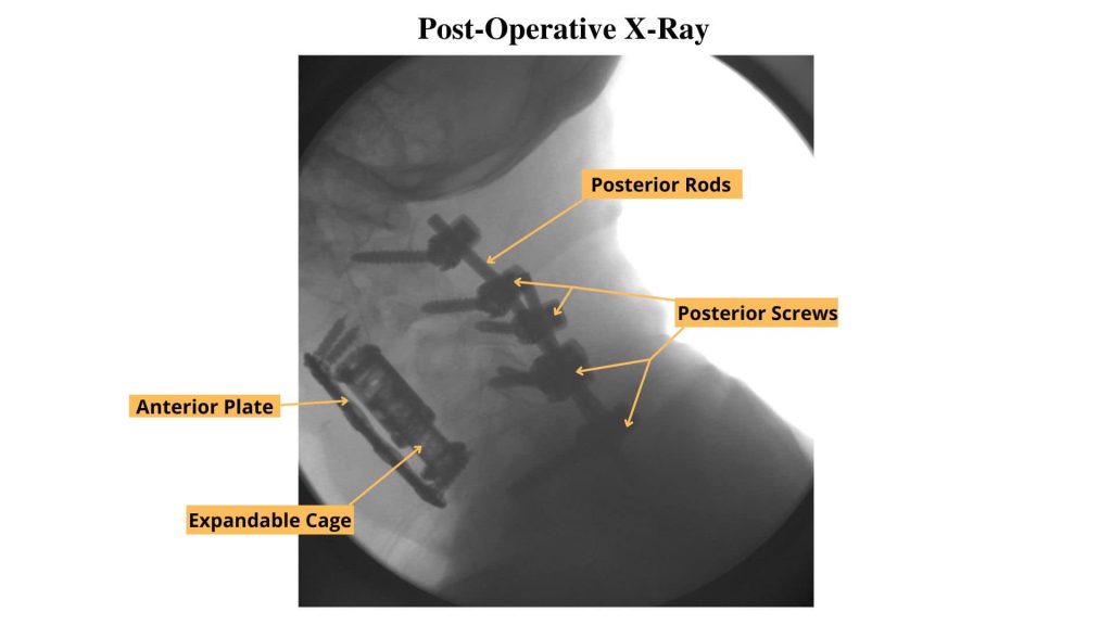
This surgery is divided into two parts: an anterior and a posterior portion. First, the patient is placed on their back to complete the anterior corpectomy and fusion. During this portion, the compressed vertebral bodies and discs of C4-6 are removed. In their place, Dr. Munshi used an expandable cage to stabilize the spine. Additionally, an anterior plate was screwed into place to ensure the spine and cage stay aligned. (Picture #2)
Following this, while the patient is still under anesthesia, the patient is placed in the prone position for the posterior portion of the surgery. This position allows for Dr. Munshi to remove bone to relieve pressure off of the spinal cord and nerve roots (Picture #2.) After successful decompression of the spinal cord, the spine is further stabilized by adding screws and rods to C2-7 (Picture #3.)
The patient tolerated the procedure well without any complications or issues. The patient was later seen in the clinic and has had significant improvements in her strength, ability to walk, and quality of life.