A 43-year-old female presented to the clinic with complaints of right lower back and buttock pain. Upon completing a physical exam and imaging, it was found that the patient had significant right sacroiliac (SI) joint osteoarthritis. She had previously had an SI joint injection which gave her good, short-term relief, however, she was interested in more long-term relief of her pain. Dr. Munshi evaluated the patient and recommended that she proceed with a right SI joint fusion.
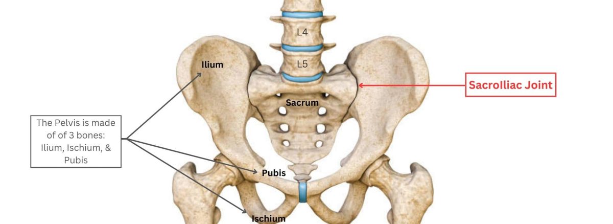
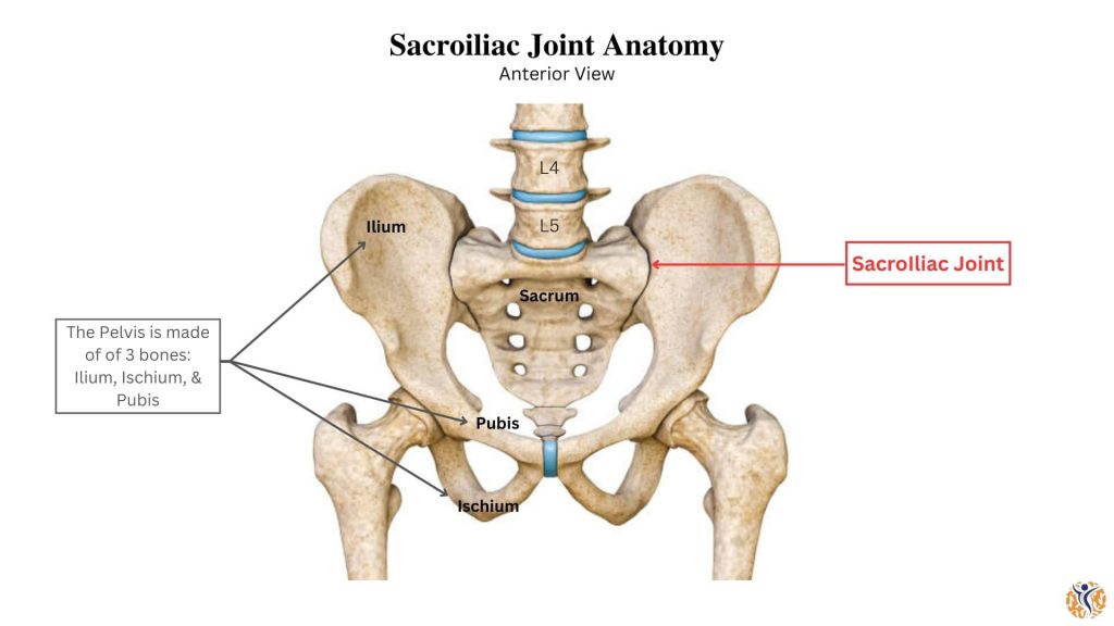
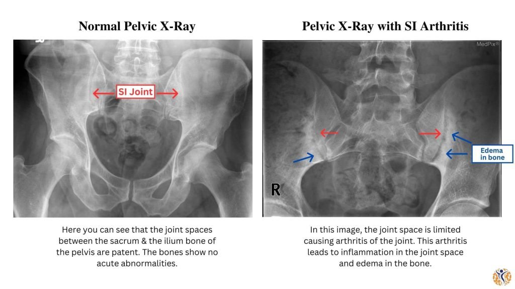
The sacroiliac, or SI joint, connects the spine at the sacrum to the pelvis at the iliac bone (Picture #1.) SI joint osteoarthritis is typically caused by degenerative changes to the joint’s cartilage (Picture #2.) These degenerative changes can be a result of injury, increased inflammation, obesity, or increasing age.
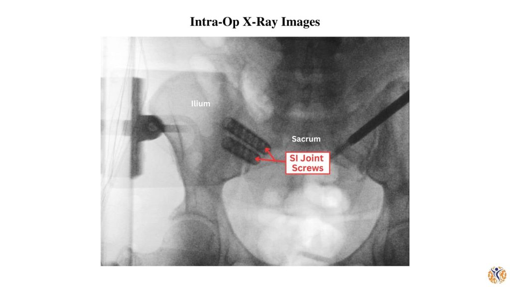
During the surgery, specialized CT-guided navigation systems are used to precisely track surgical tools and hardware. Using this specialized imaging, the sacroiliac joint is able to be fused accurately while using more minimally invasive techniques. After correctly identifying the SI joint, Dr. Munshi places screws along the joint line (Picture #3.) These screws stabilize the SI joint and reduce pain. The patient tolerated the procedure well without any complications.
Following surgery and completion of post-operative physical therapy, the patient had complete resolution of her previous pain.