A 40-year-old woman presented to the clinic with complaints of headaches, neck pain, and numbness in her bilateral upper extremities. On MRI imaging of her cervical spine, she was found to have a Chiari I malformation. Dr. Munshi evaluated the patient and recommended that she proceed with a Chiari decompression surgery for symptomatic relief.
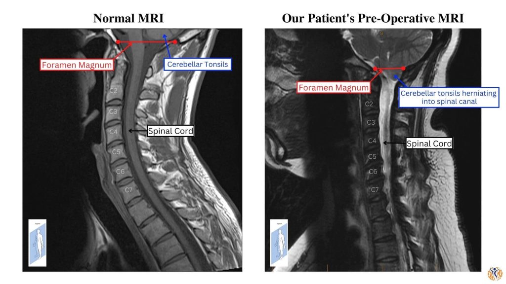
A Chiari malformation is a condition where the lower part of the brain, called the cerebellar tonsil, herniates through the skull and into the spinal canal (Picture #1.) While there are many types of Chiari malformations, type I is the most common.
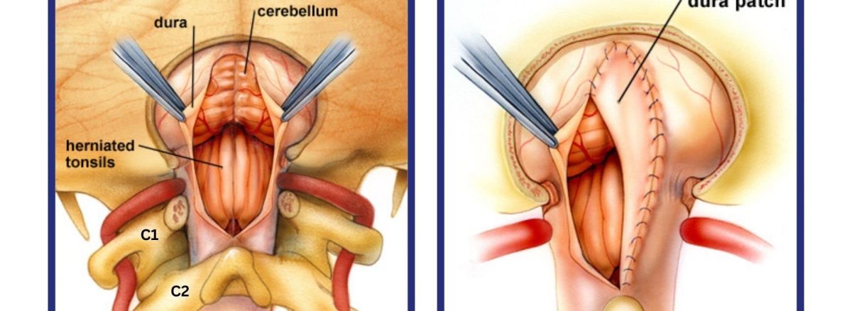
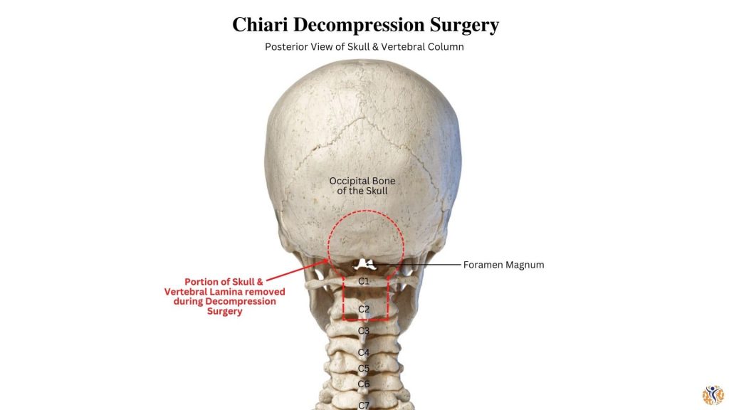
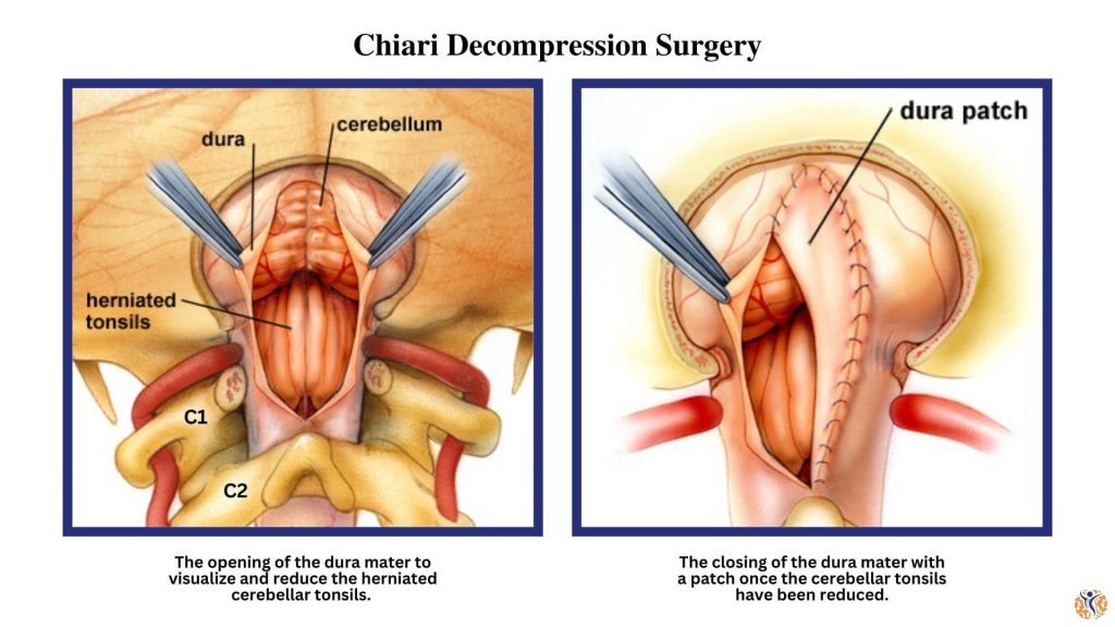
During the Chiari decompression surgery, an occipital craniotomy is performed to remove a portion of bone at the base of the skull. This widens the foramen magnum, the passage from the brain to the spinal canal, and creates space for the brain. In this case, a laminectomy of C1 and C2 was performed to completely relieve the herniated tonsils within the spinal canal (Picture #2.) Next, the dura mater, the covering of the brain, may be opened to reduce the herniated cerebellar tonsils. The dura mater is then closed with a dural patch and sealant to ensure that CSF does not leak (Picture #3.)
The patient tolerated the procedure well without any complications or issues. The patient was seen in the clinic following the surgery with improvements in her previous symptoms.