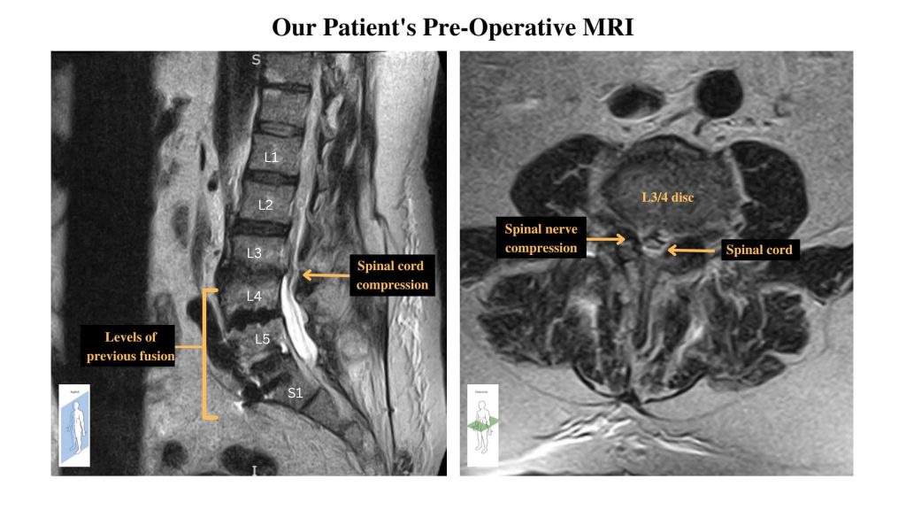
A 59-year-old man with a history of degenerative disc disease and lower back surgery, presented as a new patient to Dr. Munshi in the clinic with complaints of worsening bilateral lower back pain with radiation of pain and weakness into his left leg. His MRI imaging showed previous lumbar fusion from L4-S1 with severe canal stenosis secondary to degenerative changes at L3-4 (Picture #1.) The patient had undergone extensive conservative treatments including: activity modification, physical therapy, and epidural injections. Based on the patient’s clinical presentation and imaging, Dr. Munshi recommended that the patient proceed with an open decompression at L3-4 with an extension to the patient’s previous fusion.
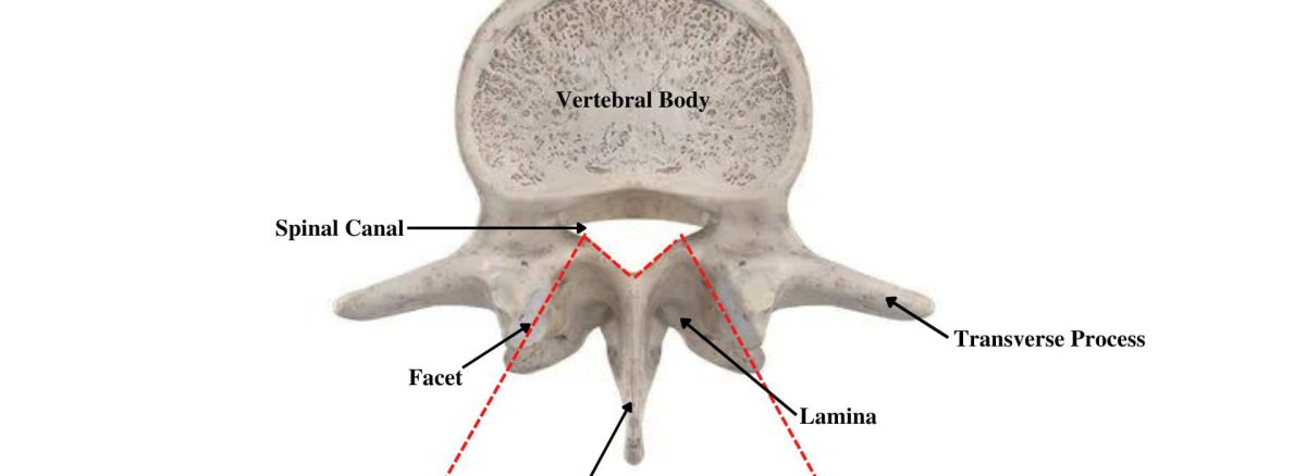
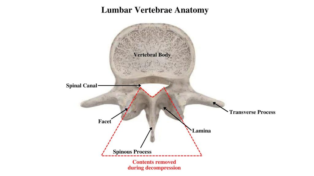
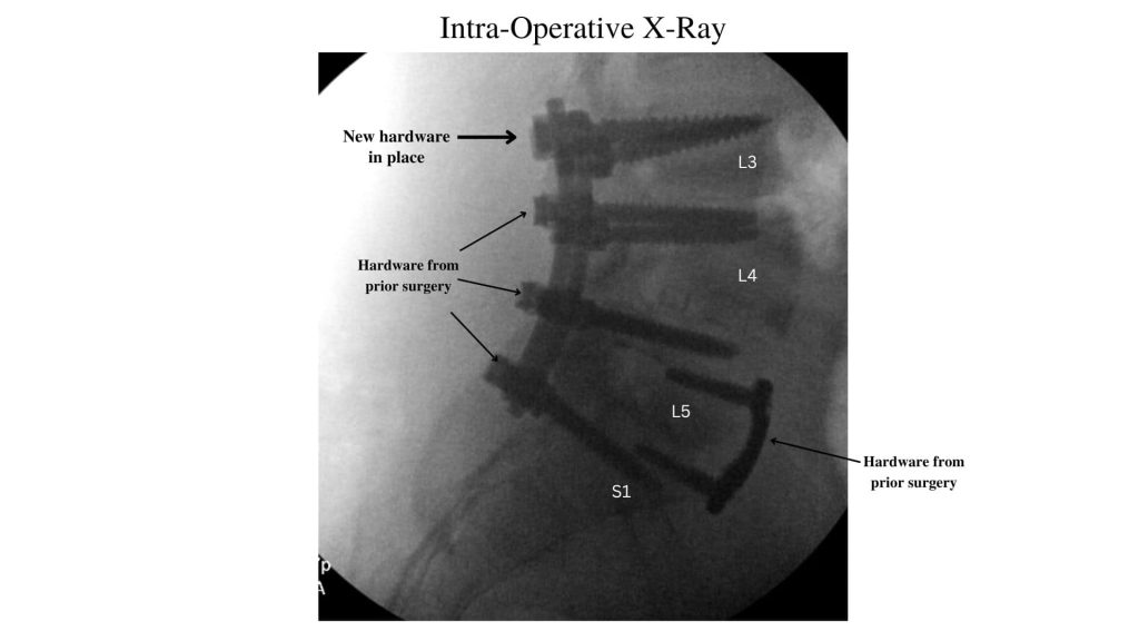
During the surgery, the pressure on the spinal cord was relieved by removing the spinous process, lamina, and part of the facet joint of the spine to relieve the pressure on the spinal cord (Picture #2.) Additionally, a foraminotomy is performed to widen the space surrounding the spinal nerve. After successful decompression at L3-4, Dr. Munshi placed screws at L3 to stabilize the decompression and new rods were added to connect to the previous fusion (Picture #3.)
The patient tolerated the procedure well without any complications. We have since seen the patient in the clinic and his lower back pain and weakness has improved.