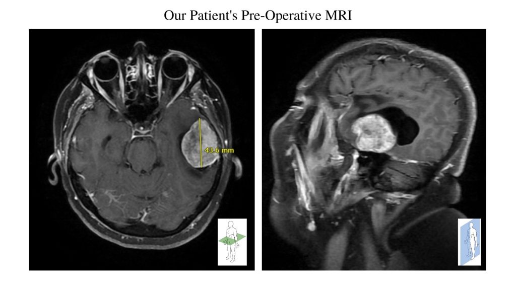
A 58-year-old female presented to the emergency department for left-sided numbness and tingling. CT and MRI imaging revealed a mass involving the left temporal and parietal lobes of the brain (Picture #1). Dr. Munshi was consulted for evaluation and determined that she would need to undergo a craniotomy for resection of the mass.
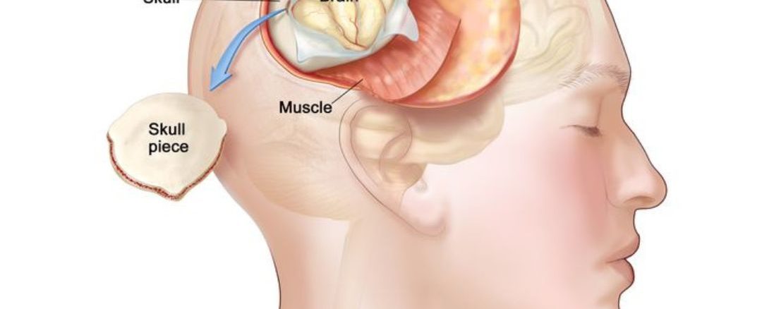
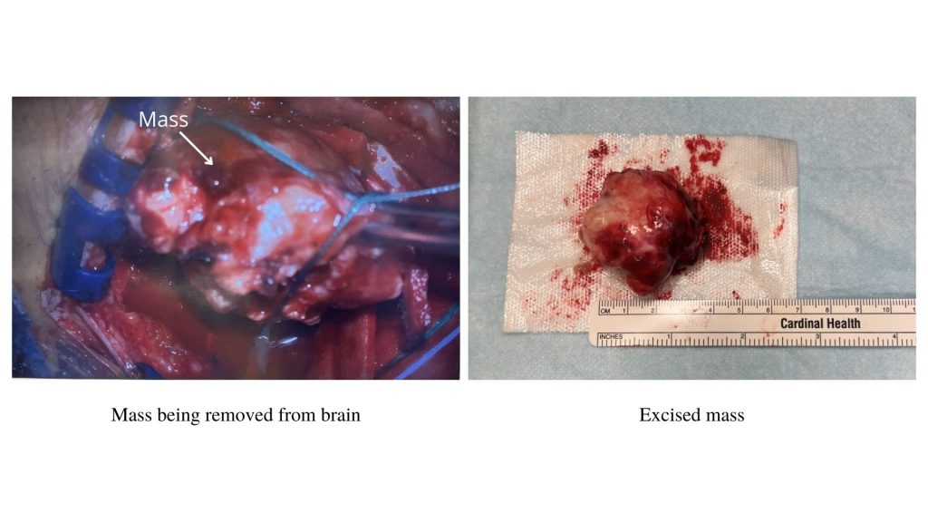
The patient underwent a left sided craniotomy, which involves removing a part of the skull to visualize the brain (Picture #2). Then, image guided navigation technology was used to pinpoint the exact location of the mass. Dr. Munshi was able to achieve complete resection of the mass during the surgery. See picture #3 for images taken during the procedure. Biopsies taken during the operative revealed the mass was benign meningioma.
A meningioma is a tumor that grows from the meninges, which are the protective membranes that cover the brain and spinal cord. Commonly, and in this case, meningiomas are slow growing and benign. Surgical intervention may be necessary for patients with large meningiomas such as this case.
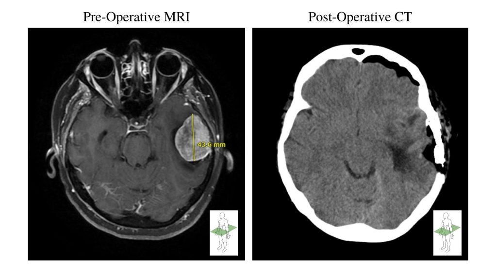
Post-operatively, a follow-up CT revealed successful resection of the mass with no complications (Picture #4).