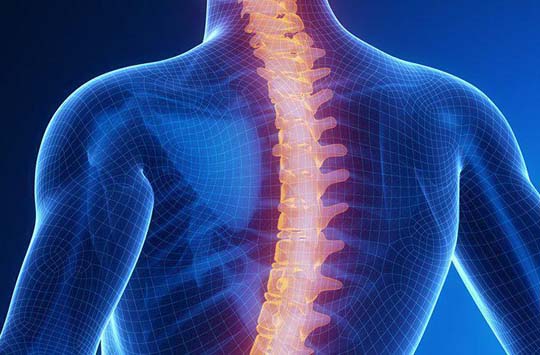
Cervical Spine Health
The cervical spine, also known as the neck, is an integral part of the human body, providing support and stability for the head and allowing for a wide range of motion. As such, it is critical to maintain cervical spine health, especially for those seeking neurological care. The cervical spine houses the spinal cord, which is responsible for transmitting signals from the brain to the rest of the body. Any damage or injury to the cervical spine can potentially cause a wide range of neurological issues, including paralysis, chronic pain, and even death.
At the Dr. Ilyas Munshi, M.D. neurological surgery clinic, maintaining cervical spine health is a top priority. With specialized expertise and advanced technologies, our neurosurgeons are equipped to diagnose and treat various cervical spine conditions, from degenerative disc disease to spinal cord tumors. By prioritizing cervical spine health, patients can improve their overall quality of life and reduce their risk of experiencing debilitating neurological complications.
Cervical Radiculopathy
Overview
Cervical radiculopathy is a problem that results when a nerve in the neck is irritated as it leaves the spinal canal.
Anatomy
The cervical spine is made up of seven vertebrae which span from the base of the skull to the thoracic spine. The vertebrae are separated by discs which help with shock absorption. The vertebrae and discs are located in front of the spinal canal where the spinal cord is located. Spinal nerves run from the spinal canal to the body through openings on both sides of the vertebrae called neural foramen. The spinal nerves are responsible for providing sensation and motor function to the areas they innervate.
In the case of cervical radiculopathy, the spinal nerve will become irritated secondary to herniated disc, osteoarthritis, or another interfering structure.
Symptoms
- Neck pain that radiates into the shoulder, arm, or the hand
- Upper extremity numbness
- Upper extremity weakness
Diagnosis
Cervical radiculopathy may be able to be diagnosed following a thorough history and physical exam. In addition to clinical findings, your healthcare provider may wish to order additional diagnostic tests to determine the cause including: x-rays, CT, or MRI imaging.
Treatment
Based on the patient’s presentation, imaging, and severity of disease initial treatment may vary. Conservative treatments include medications, heat/ice, physical therapy, and epidural injections. In some cases, surgery may be necessary.
Cervical Stenosis
Overview
Cervical stenosis is a problem that results when the spinal canal is compressed. This compression can result in irritation along the spinal cord or the spinal nerves.
Anatomy
The cervical spine is made up of seven vertebrae which span from the base of the skull to the thoracic spine. The vertebrae are separated by discs which help with shock absorption. The vertebrae and discs are located in front of the spinal canal where the spinal cord is located. Spinal nerves run from the spinal canal to the body through openings on both sides of the vertebrae called neural foramen. The spinal nerves are responsible for providing sensation and motor function to the areas they innervate.
In the case of cervical stenosis, the spinal canal which holds the spinal cord becomes compressed typically due to osteoarthritis or a disc herniation.
Causes
- Osteoarthritis/ bone spurs
- Herniated discs
- Trauma
- Spinal cord cysts or tumors
Symptoms
- Pain in neck with, or without, radiation of pain into the upper extremities
- Upper extremity numbness
- Feelings of heaviness, or weakness, in upper extremities
- Loss of functions in heads (i.e. unable to button shirt)
Diagnosis
Cervical stenosis may be able to be diagnosed following a thorough history and physical exam. In addition to clinical findings, your healthcare provider may wish to order additional diagnostic tests to determine the cause including: x-rays, CT, MRI Additionally, one might need an electromyogram (EMG) or nerve conduction studies (NCS).
Treatment
Based on the patient’s presentation, imaging, and severity of disease initial treatment may vary. Conservative treatments include medications, physical therapy, and epidural injections. In some cases, surgery may be necessary.
Cervical Disc Herniation
Overview
A cervical disc herniation occurs when an intervertebral disc tears or ruptures. The protruding disc material will cause compression and irritation to the spinal cord or nerve root.
Anatomy
The cervical spine is made up of seven vertebrae which span from the base of the skull to the thoracic spine. The vertebrae are separated by discs which help with shock absorption. The disc is made up of an inner gel-like substance which is surrounded by a fibrous cartilage. When the outer layer tears or ruptures, the gel substance can protrude, or herinate.
The vertebrae and discs are located in front of the spinal canal where the spinal cord is located. Spinal nerves run from the spinal canal to the body through openings on both sides of the vertebrae called neural foramen. The spinal nerves are responsible for providing sensation and motor function to the areas they innervate.
When the disc herniates, it can push against the spinal cord or nerve roots and cause cervical stenosis or cervical radiculopathy.
Causes
-
- Age
- Trauma
- Heavy lifting or strains
Symptoms
-
- Neck pain
- Numbness or tingling in arms and/ or hands
- Upper extremity weakness
Diagnosis
A herniated disc may be able to be diagnosed following a thorough history and physical exam. In addition to clinical findings, your healthcare provider may wish to order additional diagnostic tests to determine the cause including: x-rays, CT, CT myelogram, or MRI imaging.
Treatment
Based on the patient’s presentation, imaging, and severity of disease initial treatment may vary. Conservative treatments include medications, physical therapy, and epidural injections. In some cases, surgery may be necessary.
Cervical Degenerative Disc Disease
Overview
Degenerative disc disease is a form of arthritis which causes the discs in the spine to degenerate typically secondary to aging.
Anatomy
The cervical spine is made up of seven vertebrae which span from the base of the skull to the thoracic spine. The vertebrae are separated by discs which help with shock absorption.
The disc is made up of an inner gel-like substance which is surrounded by a fibrous cartilage. With age, typically the disc will begin to dry out and shrink. This causes the vertebrae to become closer together which may lead to the development of bone spurs.
The vertebrae and discs are located in front of the spinal canal where the spinal cord is located. Spinal nerves run from the spinal canal to the body through openings on both sides of the vertebrae called neural foramen. The spinal nerves are responsible for providing sensation and motor function to the areas they innervate.
Causes
- Age
- Excessive straining
- Obesity
Symptoms
-
- Neck and/ or shoulder pain
- Upper extremity pain
- Upper extremity numbness
Diagnosis
Degenerative disc disease may be able to be diagnosed following a thorough history and physical exam. In addition to clinical findings, your healthcare provider may wish to order additional diagnostic tests to determine the cause including: x-rays, CT, or MRI
Treatment
Based on the patient’s presentation, imaging, and severity of disease initial treatment may vary. Conservative treatments include medications, physical therapy, and epidural injections. In some cases, surgery may be necessary.
Anterior Cervical Discectomy and Fusion (ACDF)
Overview
Anterior cervical discectomy and fusion (ACDF) is a neck surgery that has 2 main components…
-
- A cervical discectomy which involves the removal of disc between two vertebrae
- A fusion, which is performed at the same time as the discectomy, which stabilizes the cervical spine. The fusion may be performed with bone grafts and/or implantable hardware
An ACDF may be done at a single level, or multiple levels depending on the patient
Who is a Candidate?
This surgery is typically performed for symptomatic relief of a cervical herniated disc, degenerative disc disease, or cervical spinal stenosis. Typically surgery may be an option if physical therapy or medications fail to relieve symptoms.
What Happens During Surgery?
-
- A small horizontal skin incision is made on the anterior aspect of the neck.
- Next, neck muscle, esophagus, trachea, and carotid arteries are retracted to visualize the anterior aspect of the spine.
- Specialized x-ray imaging, called fluoroscopy, is used during the procedure to identify the correct vertebral and disc levels.
- Once the surgeon has identified the correct spinal level, the appropriate discs are removed.
- Following the disc removal, typically the surgeon will also remove excess bone, or osteophytes, ensuring that there is no excess material that can cause irritation to the spinal cord or nerve roots. This is often done with the assistance of a microscope.
- Next, the fusion of the cervical segment is done to ensure stabilization of the vertebrae. The fusion may be performed with bone grafts and/or implantable hardware.
- In addition to the fusion between vertebrae, the surgeon may opt to add a small plate to the anterior aspect of the vertebrae. This provides additional stability.
- The muscle and skin is then closed with sutures and a gauze dressing is placed on the skin.
Recovery
-
- Most patient typically stay 1 night in the hospital following the surgery
- Immediately following surgery, the patient will be unable to lift anything over 5 pounds, bend over completely, twist, or drive until they follow-up in clinic, approximately 2-3 weeks after surgery
- Most patients recover and return to most everyday activities 4-6 weeks following surgery
Prevention
-
- Recurrences in neck pain are common. In order to prevent recurrence of cervical pain it is important to maintain the following…
- Proper lifting techniques
- Good posture while sitting, standing, moving, and sleeping
- Appropriate exercise
- Ergonomic work areas
- Healthy weight
- No smoking
Potential Complications
-
- Inadequate symptom relief
- Temporary or persistent difficulty swallowing
- Hardware failure
- Nerve root damage
- Spinal cord damage
- No surgery is risk free, other complications include, but are not limited to, bleeding, infection, injury, or even death.
Disclaimer: This information is strictly informational and not intended for medical advice. If you have any questions about surgical procedures, symptoms, or restrictions following surgery please contact your physician.
Posterior Cervical Decompression and Fusion (PCDF)
Overview
A posterior cervical decompression and fusion (PCDF) occurs in the back of the neck to relieve pressure on the nerves or spinal cord.
Who is a Candidate?
This surgery is typically performed for symptomatic relief of a cervical herniated disc, cervical kyphosis, or cervical spinal stenosis. Typically surgery may be an option if physical therapy or medications fail to relieve symptoms.
What Happens During surgery?
-
- A vertical skin incision is made on the posterior aspect of the neck.
- Next, specialized x-ray imaging, called fluoroscopy, is used during the procedure to identify the correct vertebral and disc levels.
- To achieve successful decompression, typically the surgeon will remove herniated disc material and excess bone to ensure that there is no excess material that can cause irritation to the spinal cord or nerve roots. This may be done with the assistance of a microscope.
- Next, the fusion of the cervical segment is done to ensure stabilization of the vertebrae. The fusion may be performed with bone grafts and/or implantable hardware.
- The muscle and skin is then closed with sutures and a gauze dressing is placed on the skin.
Recovery
-
- Most patient typically stay 1-2 nights in the hospital following the surgery
- Immediately following surgery, the patient will be unable to lift anything over 5 pounds, bend over completely, twist, or drive until they follow-up in clinic, approximately 2-3 weeks after surgery
- Most patients recover and return to most everyday activities 3-6 months following surgery
Prevention
Recurrences in neck pain are common. In order to prevent recurrence of cervical pain it is important to maintain the following…
-
- Proper lifting techniques
- Good posture while sitting, standing, moving, and sleeping
- Appropriate exercise
- Ergonomic work areas
- Healthy weight
- No smoking
Potential Complications
-
- Inadequate symptom relief
- Hardware failure
- Nerve root damage
- Spinal cord damage
- No surgery is risk free, other complications include, but are not limited to, bleeding, infection, injury, or even death.
Disclaimer: This information is strictly informational and not intended for medical advice. If you have any questions about surgical procedures, symptoms, or restrictions following surgery please contact your physician.