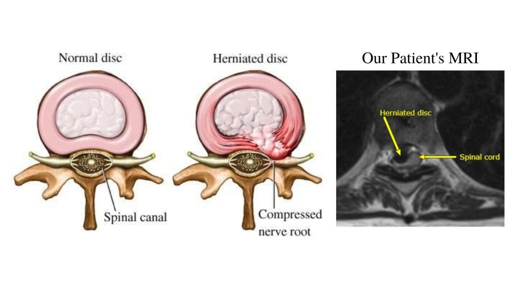
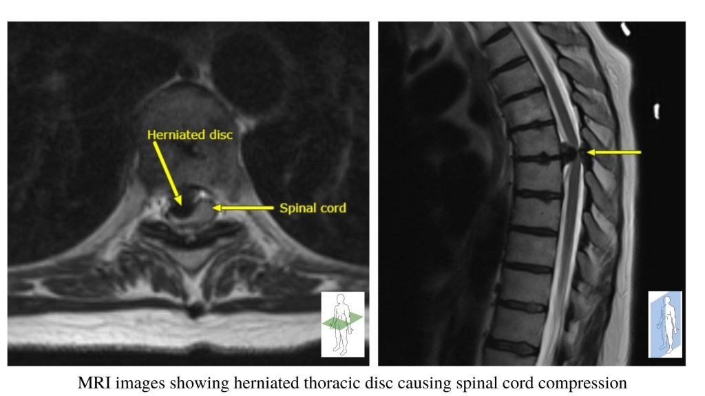
A 54-year-old woman presented to the clinic with complaints of worsening left sided weakness and numbness. On MRI, she was found to have a large herniated disc extrusion at T6/7 which was severely compressing the thoracic spinal cord.
A herniated thoracic disc is a spinal condition in which the soft center of the intervertebral disc (the nucleus pulposus) pushes through a tear in the tough outer layer of the disc (the annulus fibrosus) and into the spinal canal, causing compression on the spinal canal and/ or spinal nerve. Picture #1 & #2 show examples of herniated discs, as well as the patient’s actual MRI images.
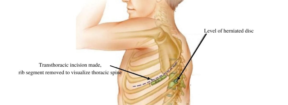
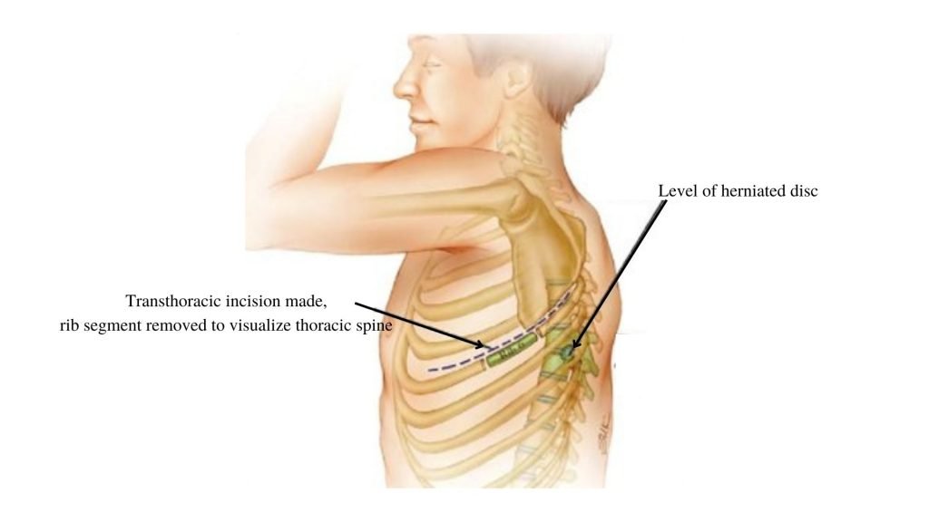
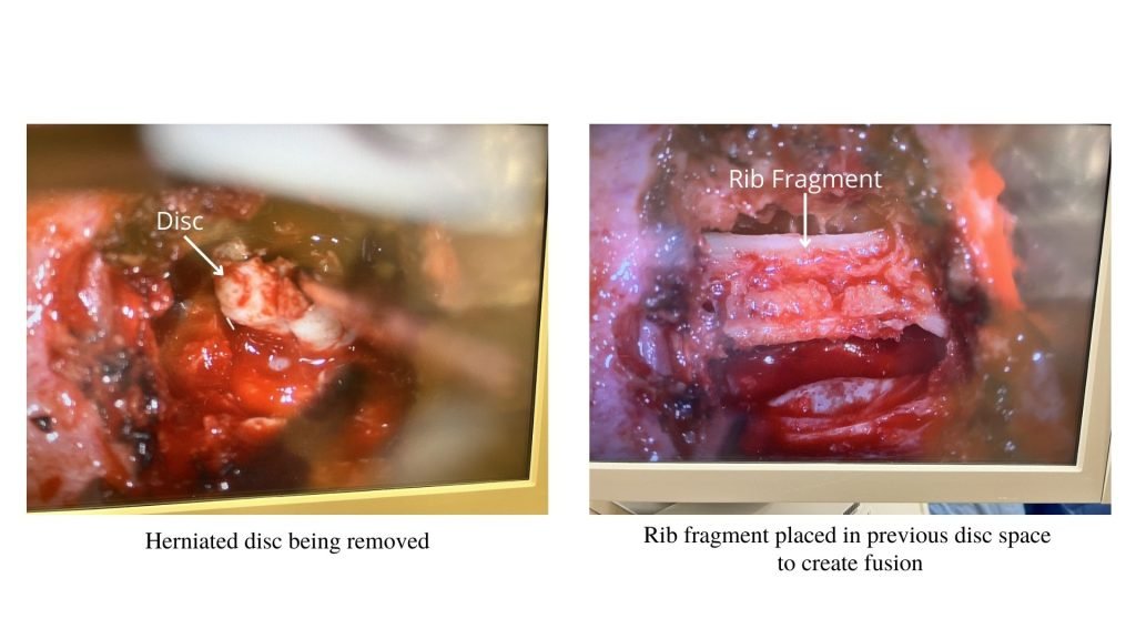
In this case, the patient was placed on their side and underwent a transthoracic removal of rib with a subsequent anterior lateral discectomy to remove the herniated disc (Picture #3, #4) Following the removal of the damaged disc, the patient’s own rib was used to create a fusion to stabilize the adjacent vertebrae (Picture #4.) At the end of the procedure, a chest tube was left in place to remove excess air in the thoracic cavity. The chest tube was monitored during their hospital stay and subsequently removed with no complications.
The patient tolerated the procedure well without and since the patient’s weakness and numbness has significantly improved.